Diagnostic Nuclear Medicine
What is Diagnostic Nuclear Medicine
In diagnostic nuclear medicine, the unique characteristics of chemicals called radio-pharmaceuticals* are used for diagnosis. Typically, a small amount of a radiopharmaceutical is introduced into the body by injection, ingestion, or inhalation. The radiopharmaceutical is attracted to particular organs, bones, or other tissues. The radioisotope releases small amounts of energy (radiation) that can be detected outside the body by special "cameras". These cameras record the movement and localization of radiopharmaceuticals in the body. The resulting 2- and 3-dimensional images document the function (metabolic, physiologic, and pathologic) of the tissue or organ of interest. Physicians examine these images to evaluate and diagnose a large number of diseases.
Some procedures in nuclear medicine do not involve imaging. These tests involve the injection of a small amount of radiopharmaceutical usually followed by the taking of blood samples for analysis.
* Radiopharmaceuticals are molecules or chemicals that are attached to a small amount of radioactive isotope that once administered to the patient are able to specifically localize within organs and/or organ systems in health and disease.
Some common procedures
Nuclear medicine images can assist your doctor in diagnosing diseases or planning and monitoring your treatment. Tumours, infection and other disorders can be detected by evaluating organ function.
The most common procedures carried out are:
- Bone Scan - to evaluate bones for fracture, infection, arthritis or tumour, or determine the presence or spread of cancer
- Lung scan - to look at respiratory and blood-flow problems
- Myocardial perfusion scan (MPS scan) - to image blood flow and function of the heart
- Multi gated cardiac scan (MUGA scan) - to image blood flow and function of the heart
- Renal static scan (DMSA scan) - to locate the presence of infection
- Renal dynamic scan (MAG3 or DTPA scan) - to analyze kidney function
- Sentinel lymph node scan (SLN scan)
- Iodine whole body scan
- Glumerular filtration rate measurement (GFR)
- Thyroid uptake measurement - to measure thyroid function to detect an overactive or underactive thyroid
- Other scans
What does the equipment look like?
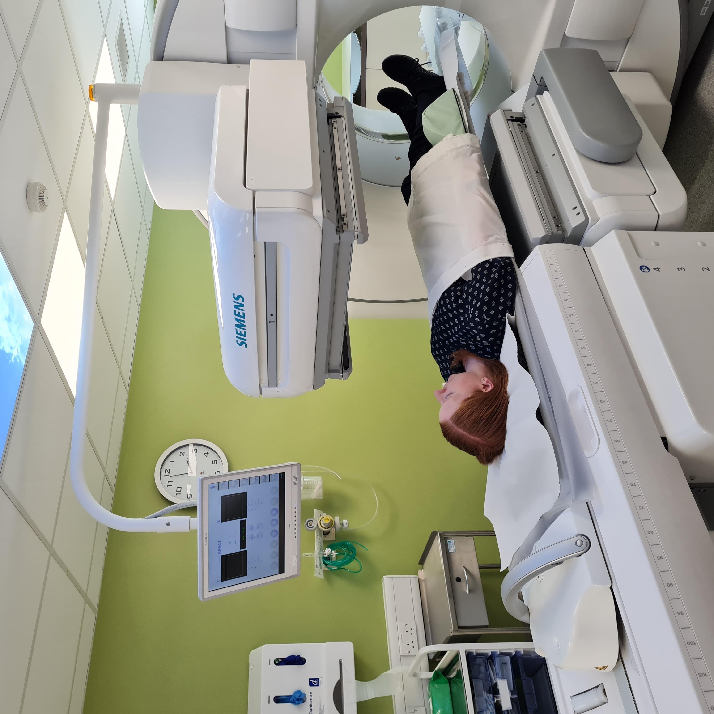
For imaging procedures we use a gamma camera. This is a piece of equipment which can detect radiation and from where it is coming. During most nuclear medicine examinations, you will lie down on a scanning table. The gamma camera will take pictures of the parts of your body in which the doctors are interested. Some procedures involve scanning your whole body. The images are stored on a computer for processing.

How is an imaging procedure performed?
Depending on the type of scan, it may take several seconds to several days for the substance to travel through the body and accumulate in the organ to be studied, thus there is a wide range in scanning times. While the images are being obtained, you must remain as still as possible. This is especially true when a series of images is obtained to show how an organ functions over time or when a tomographic* procedure is taking place. After the procedure, the radiographer will check that the quality of the images are optimal for processing and diagnosis. When this is done you will be able to leave. The images will then be processed to enable a doctor with specialised training in nuclear medicine to report on the findings and let your doctor know within a few days.
* A tomographic image is created by letting the gamma camera rotate around your body.
What will I experience during the procedure?
Some minor discomfort during a nuclear medicine procedure may arise from the intravenous injection, usually done with a small needle. Lying still on the examining table may be uncomfortable for some patients. The amount of radiopharmaceutical we use is very small and you would normally not feel anything from this. During the scan you should be as still as possible to enable us to collect good images at the first attempt.
For cardiac procedures we will sometimes need to get your heart to beat faster than normal and we will use a special drug to achieve this. Patients normally experience some minor symptoms from this drug, but they will quickly go away once we stop giving this drug. You will be monitored by highly qualified staff while this is taking place.
If you are having a non-imaging test you can leave between the injection and the blood sampling if you so wish. Just make sure you return for the blood sampling at the correct time.
Who will interpret the results and how do I get them?
Most patients undergo a nuclear medicine examination because their primary care doctor has recommended it. A physician who has specialised training in nuclear medicine will interpret the images or test results and forward a report to your doctor. It usually takes a few days to interpret, report and deliver the results.
What are the benefits versus risks
Benefits:
- The functional information provided by nuclear medicine examinations is unique and can not be obtained by using other procedures. For many diseases, nuclear medicine studies will give the most useful information needed to make a diagnosis and to determine appropriate treatment, if any.
- Nuclear medicine is much less traumatic than exploratory surgery, and allergic reaction to the radiopharmaceutical material is extremely rare.
Risks:
- Because the doses of radiopharmaceutical administered are very small, nuclear medicine procedures result in exposure to a small dose of radiation. Nuclear medicine has been used for more than five decades, and there are no known long-term adverse effects from such low-dose studies. The dose you will receive is similar to an X-ray examination and can be compared to the dose you receive from natural background radiation* over a few days to a couple of years, dependent on the type of procedure you have.
- As with all radiologic procedures, be sure to inform your physician if you are pregnant. In general, exposure to radiation during pregnancy should be kept to a minimum.
- Allergic reactions to the radiopharmaceutical can occur, but are extremely rare.
*Everybody is exposed to what is called natural background radiation coming from space, building material the ground, e.g. radon gas and from radioactivity inside our body. This natural radiation gives us a radiation dose equivalent to many hundred chest X-rays a year.
What are the limitations of general nuclear medicine?
Nuclear Medicine procedures are time-consuming. They involve administration of a radiopharmaceutical, acquisition of images or analysis of blood or urine samples and interpretation of the results. It can take hours to days for the radiopharmaceutical to accumulate in the part of the body under study. Imaging and some non-imaging procedures can take up to three hours to perform.


