
Clinical Advice
This is available on a 24/7 basis by contacting the on-call Haematology SpR or Consultant Haematologist via the Trust Switchboard 01202 303626
Investigation of Bleeding Disorders
Patients with excessive bruising or bleeding should be investigated initially with a full blood count and coagulation screen, giving any clinical details of family history of bleeding, and patients bleeding after surgery, dental extraction etc.
Following these initial screening tests, further appropriate investigation (e.g. for Von Willebrand’s Disease) will be advised and arranged by a Consultant Haematologist either via referral or A&G on ERS.
Known Patients with Bleeding Disorders
Local patients with known bleeding disorders are registered with, and followed up by, the Haemophilia Centre at University Hospitals Dorset.
These patients have open access for bleeding problems
Dr Luke Attwell, Consultant Haematologist, Royal Bournemouth Hospital is the Director of the Haemophilia Service in Dorset
All patients should carry a red bleeding disorder card which contains information about the specific coagulation factor levels and emergency telephone numbers. (Holiday visitors with bleeding disorders should also carry these cards, which should provide the necessary information)
D-Dimers
Following recent enquiries from GPs concerning D-Dimers, Dr Luke Attwell has reviewed the procedure for telephoning D-Dimer results and has prepared the following information:
D-Dimers are breakdown products of cross-linked fibrin, and a marker for increased fibrinolysis
Cause of raised D-Dimers
- Acute thrombosis
- Cancer
- Diabetic ketoacidosis
- Disseminated intravascular coagulation (DIC)
- Infection
- Pregnancy
- Surgery
- Trauma
Normal range
0 – 0.5 µg/mL based on the manufacturer’s recommendations. An age adjusted D-dimer is now used. If aged 51 years or older the upper limit of the normal range is calculated by (age ÷ 100). This is reflected on the individual patient reference range.
Telephoning D-Dimer results
GP requests for D-Dimers will only be telephoned to the requestor when they exceed a value of 1 µg/mL
D-Dimers in the diagnosis of a VTE
The Negative Predictive Value is 99% if used in conjunction with a negative clinical probability score (such as Wells)
It would be a very rare event to have a patient with a venous thromboembolism (VTE) when the D-Dimer is < 0.5 µg/mL
The importance of performing a Wells score cannot be understated
A patient with a normal D-Dimer and low probability Wells score is highly unlikely to have a VTE
A D-Dimer is not required when a patient has a high probability of a VTE such as a Wells score of > 2 in the context of a possible deep venous thrombosis or > 4 in the context of a possible pulmonary embolus or if a patient is taking anticoagulation therapy
These patients will require a scan to confirm or exclude VTE
D-Dimers can return to normal within 4 weeks of suffering a VTE and after starting anticoagulation therapy
D-Dimer production is known to be suppressed in patients receiving anticoagulation therapy e.g. Warfarin, so their measurement can be unreliable in trying to exclude VTE in this context and may be misleading.
For clinical queries contact Dr Luke Attwell – Consultant Haematologist
Thrombophilia Guidelines
Guidance
For most unselected individuals in the general population without a personal history of VTE, the risk-benefit analysis does not favour testing for hereditary thrombophilia. Please see trust guidelines for further advice.
A thrombophilia screen includes the following testing: antithrombin, protein C & S levels, factor V Leiden mutation, prothrombin gene mutation and antiphospholipid testing.
Consider thrombophilia testing for the following indications:
- Patients presenting with venous thrombosis who are < 40 years old and do not have a precipitating cause and are > 3 months from diagnosis of VTE
- Patients presenting with an unprovoked venous thrombosis who have an apparent thrombosis-prone family (more than one symptomatic family member) and are > 3 months from diagnosis of VTE
- Patients with a first-degree relative with confirmed inherited thrombophilia
- Patients presenting with a venous thrombosis at an unusual site without an obvious cause (e.g. mesenteric thrombus or cerebral venous sinus thrombosis) consider testing for APS, MPN and PNH
- Antiphospholipid antibodies and lupus anticoagulant testing in patients < 50 years old with a stroke and no identified risk factors for arterial disease or in those with recurrent events despite best medical therapy and no other identified causes
- Consider MPN and PNH testing for arterial stroke if FBC results are suggestive.
- Antiphospholipid testing in patients with recurrent pregnancy-related morbidity e.g. recurrent miscarriage.
The genetic mutations screened as part of the thrombophilia investigation are Factor V Leiden G1691A and Prothrombin Gene G20210A
Malaria
All patients who have a history of travel to or through (including an airport “stop-over”) an area where malaria is endemic, and who present to a general practitioner unwell, should immediately have a blood film checked for malarial parasites.
Patients in whom malaria is suspected should be referred to the Medical Registrar on duty in the Emergency Department immediately. Please indicate on the request form the malarial area of the world visited
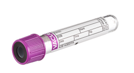
Blood samples should be sent in a Full Blood Count tube (Purple cap)
Further films should be inspected if parasites are not found on initial screening and the patient remains unwell
Immediate and appropriate treatment may be lifesaving in Plasmodium falciparum (malignant tertian) malaria
Guidance for patients with polycythaemia
The investigation pathway for patients with increased haematocrit in primary and secondary care is given below as a guide
JAK2 should only be requested via the GP if suggested by Haematology as part of Advice and Guidance as the patient should be referred to the Clinical Haematology team if the result is positive
Hct >0.56 (Male), Hct >0.54 (Female) Causes of secondary (including apparent) polycythaemia include hypoxia, for example in chronic lung disease, smoking, diuretic use, androgens, rarely erythropoietin producing tumours. Please repeat, ensuring that the patient is well-hydrated. If persistent and unexplained, please check MPN screen and serum erythropoietin level and discuss with haematology.
Sickle Cell Disease Adult Guidelines
Haematology at UHD links with the Wessex & Thames Valley Haemoglobinopathy Coordinating Centre
Guidelines can be found via this link: NSSG Haematology – Adult Haemoglobinopathies
Guideline for the Investigation of a Paraprotein
The presence of a paraprotein is common and increases with age. In many cases no underlying cause for the paraprotein will be found. These cases can be labelled as MGUS (monoclonal gammopathy of uncertain significance). However, the presence of a paraprotein may represent more serious conditions such Multiple Myeloma (MM), Waldenstrom’s macroglobulinaemia (WM), Amyloidosis or other types of lymphoma.
In patients who have features suggestive of one of these conditions, full staging including bone marrow biopsy is indicated. However, in an elderly patient group it is often inappropriate to perform full staging investigations in all patients.
Studies have shown that the likelihood of myeloma is small if the paraprotein level is low. In addition, it is well established that there is no long-term benefit in treating such conditions as MM and WM early before symptoms of the condition or investigative evidence of end organ damage occurs.
The finding of acquired hypogammaglobulinaemia (immuneparesis) without the presence of a paraprotein should also be investigated in the same way as a paraprotein.
- Initial investigation of all paraproteins (IgG, A or M) should include:
- Clinical history specifically to identify bone pain (usually back), recurrent infection, symptoms of hyperviscosity, symptoms of anaemia, symptoms of hypercalcaemia and peripheral neuropathy.
- Patient examination for evidence of anaemia, lymphadenopathy, hepatosplenomegaly, neuropathy and hyperviscosity (fundi).
- FBC, CRP, renal, liver and bone profiles.
- Serum free light chain ratio, LDH and b2microglobulin.
- Further management of an IgG or IgA monoclonal paraprotein:
- Patients with a low level paraprotein: IgG < 15g/l and IgA < 10g/l who have no evidence of immuneparesis i.e. other immunoglobin levels are normal.
And
- Have a normal serum free light chain ratio (or a kappa/lambda excess <200)
And
- NO evidence of end organ damage:
- Calcium normal
- Renal function normal
- No anaemia or other cytopenia
And
- NO symptoms suspicious of underlying myeloma (bone pain, recurrent infection etc.)
If ALL the above criteria are met, these patients do not require further investigation but should be followed up with paraprotein measurement every 6 months initially.
If after 2 measurements the paraprotein level is stable blood measurements can be reduced to annually.
Indications for haematological referral for further investigations:
- A paraprotein is found and the patient does not meet the above criteria for monitoring in primary care.
- A low level paraprotein is found but evidence of immuneparesis or suspicious clinical symptoms or anaemia, hypercalcaemia or renal impairment are present.
- Further management of an IgM monoclonal paraprotein:
Patients with a low level paraprotein: IgM < 10g/l who have no evidence of immuneparesis i.e. other immunoglobin levels are normal.
And
- NO evidence of end organ damage:
- Calcium normal
- Renal function normal
- No anaemia, other cytopenia or lymphocytosis
And
- NO symptoms attributable to the IgM paraprotein or suspicious of underlying lymphoproliferative disease (recurrent infection, lymphadenopathy, hepatosplenomegaly, peripheral neuropathy, cold agglutinin disease, hyperviscosity symptoms).
If ALL the above criteria are met, these patients do not require further investigation but should be followed up with paraprotein measurement every 6 months initially.
If after 2 measurements the paraprotein level is stable blood measurements can be reduced to annually.
Indications for haematological referral for further investigations:
- An increase in paraprotein to above the stated levels or any other change resulting in the patient no longer meeting the above criteria for observation only.
- A low level paraprotein is found but evidence of immuneparesis or suspicious clinical symptoms or cytopenia/lymphocytosis etc. are present.
NOTE: Patients with a significant paraprotein can be referred by a GP via the ‘fast-track’ system if they additionally have any of the following;
- Persistant lymphadenopathy
- Hepatosplenomegaly
- Bone pain with anaemia and raised CRP
- X-rays with lytic lesions
- Three or more of fatigue, recurrent infections, weight loss, bone pain, bruising, breathlessness, itching.
For further information contact Haematology secretaries, 0300 019 4790.
Test Repertoire and Reference Ranges
Test repertoire can be found on the Blood Sciences Test Guide
Select ‘Haematology’ discipline and use the A to Z or search function.
Reference Ranges:
Reference ranges for Haematology & Coagulation (Adult, Paediatric and Infant):
Adult FBC Reference Ranges
|
|
Reference Range | |
|---|---|---|
|
Parameter |
Male |
Female |
|
Red Blood Cells (RBC) x 1012/L |
4.5 - 5.5 |
3.8 – 4.8 |
|
Haemoglobin (HB) g/L |
130 - 170 |
120- 150 |
|
Haematocrit (Hct) |
0.4 - 0.5 |
0.36 - 0.46 |
|
Mean Cell Volume (MCV) fL |
83 - 101 |
|
|
Mean Cell Haemoglobin (MCH) pg |
27 - 32 |
|
|
Mean Cell Haemoglobin Concentration (MCHC) g/L |
315 - 345 |
|
|
Platelets (PLT) x 109/L |
150 - 410 |
|
|
White Blood Cells (WBC) x 109/L |
4.0 – 10.0 |
|
|
Differential: x 109/L Neutrophils Lymphocytes Monocytes Eosinophils Basophils |
2.0 ‑ 7.0 (40-80%) 1.0 - 3.0 (20-40%) 0.2 ‑ 1.0 (2-10%) 0.02 ‑ 0.5 (1-6%) 0.02 ‑ 0.1 (<1-2%) |
|
|
Reticulocytes x 109/L |
50 – 100 |
|
Paediatric Reference Ranges
|
|
Reference Range | ||
|---|---|---|---|
|
Parameter |
1 year |
2 - 6 Years |
6 – 12 Years |
|
RBC (x 1012/L) |
3.9 - 5.1 |
4.0 - 5.2 |
4.0 - 5.2 |
|
HB (g/L) |
111 - 141 |
110-140 |
115 - 155 |
|
Hct |
0.30 - 0.38 |
0.34 - 0.40 |
0.35 - 0.45 |
|
MCV (fL) |
72 – 84 |
75 - 87 |
77 - 95 |
|
MCH (pg) |
25 – 29 |
24 – 30 |
25 – 33 |
|
MCHC (g/L) |
320 – 360 |
310 – 370 |
310 – 370 |
|
PLT (x 109/L) |
200 - 550 |
200 - 490 |
170-450 |
|
WBC (x 109/L) |
6 - 16 |
5 - 15 |
5 - 13 |
|
Differential: Neutrophils Lymphocytes Monocytes Eosinophils (x 109/L) |
1.0 ‑ 7.0 3.5 – 11.0 0.2 ‑ 1.0 0.1 – 1.0 |
1.5 ‑ 8.0 6.0 – 9.0 0.2 ‑ 1.0 0.1 – 1.0 |
2.0 ‑ 8.0 1.0 – 5.0 0.2 ‑ 1.0 0.1 – 1.0 |
|
Reticulocytes (x 109/L) |
30 - 100 |
30 - 100 |
30 - 100 |
Infant Reference Ranges
|
|
Reference Range | ||||||
|---|---|---|---|---|---|---|---|
| Parameter |
BIRTH |
DAY 3 |
DAY 7 |
DAY 14 |
1 MONTH |
2 MONTHS |
3-6 MONTHS |
|
RBC (x1012/L) |
5.0 - 7.0 |
4.0 - 6.6 |
3.9 - 6.3 |
3.6 - 6.2 |
3.0 - 5.4 |
3.1 - 4.3 |
4.1 – 5.3 |
|
Hb (g/L) |
140 - 220 |
150 - 210 |
135 - 215 |
125 - 205 |
115 - 165 |
94 - 130 |
111 - 141 |
|
Hct |
0.45 - 0.75 |
0.45 - 0.67 |
0.42 - 0.66 |
0.31 - 0.71 |
0.33 - 0.53 |
0.28 - 0.42 |
0.3 – 0.4 |
|
MCV (fL) |
100 - 120 |
92 – 118 |
88 - 126 |
86 – 124 |
92 - 116 |
87 - 103 |
68 - 84 |
|
MCH (pg) |
31 - 37 |
31 - 37 |
31 - 37 |
31 – 37 |
30 - 36 |
27 - 33 |
24 - 30 |
|
MCHC (g/L) |
300 - 360 |
290 - 370 |
280 - 380 |
280 – 380 |
290 - 370 |
285 - 355 |
300 - 360 |
|
PLT (x 109/L) |
100 - 450 |
210 – 500 |
160- 500 |
170 – 500 |
200 - 500 |
210 - 650 |
200 - 550 |
|
WBC (x 109/L) |
10 - 26 |
7 - 23 |
6 - 22 |
6 – 22 |
5 – 19 |
5 - 15 |
6 -18 |
|
Neutrophils Lymphocytes Monocytes Eosinophils (x 109/L) |
4 ‑ 14 3 - 8 0.5 ‑ 2.0 0.1 - 1.0 |
3 ‑ 5 2 - 8 0.5 ‑ 1.0 0.1 - 2.0 |
3 - 6 3 - 9 0.1 - 1.7 0.1 - 0.8 |
3 ‑ 7 3 - 9 0.1 ‑ 1.7 0.1 - 0.9 |
3 ‑ 9 3 - 16 0.3 ‑ 1.0 0.2 - 1.0 |
1 ‑ 5 4 - 10 0.4 ‑ 1.2 0.1 - 1.0 |
1 – 6 4 –12 0.2 – 1.2 0.1 - 1.0 |
|
Reticulocytes (x 109/L) |
120 - 400 |
50-350 |
50 - 100 |
50 - 100 |
20 - 60 |
30 - 50 |
40 - 100 |
ESR Reference ranges:
|
ESR (mm in 1hr) Starrsed EDTA method | |
|---|---|
|
Male 0 – 50 years |
1-10 |
|
Female 0 – 50 years |
1-12 |
|
Male 51-60 years |
1-12 |
|
Female 51-60 years |
1-19 |
|
Male 61-70 years |
1-14 |
|
Female 61-70 years |
1-20 |
|
Male > 70 years |
1-30 |
|
Female > 70 years |
1-35 |
Manual ESR reference Range:
Performed on patients <10 years old
|
ESR (mm in 1hr) citrate manual method Polymedco Sediplast | |
|---|---|
|
Male 0 – 50 years |
1-8 |
|
Female 0 – 50 years |
1-10 |
|
Male 51-70 years |
1-12 |
|
Female 51-70 years |
1-17 |
|
Male > 70 years |
1-25 |
|
Female > 70 years |
1-29 |
Haematology FBC and ESR Reference Ranges are taken from Dacie & Lewis 12th edition
Tosoh G11 HbA1c Reference Range:
|
HbA1c (mmol HbA1c/mol HbA) IFCC Non-Diabetic Normal Range |
25-41 |
Stago STAR MAX 3 Coagulation Screen and D-Dimer Reference Ranges:
Adult Reference Ranges (10 years and above)
|
Parameter |
Reference Range |
|---|---|
|
Prothrombin Time (in Seconds) |
11.6-14.6 |
|
International Ratio (INR) |
0.9-1.2 |
|
Activated Partial Thromboplastin Time (APTT) (in Seconds) |
27.0-37.3 |
|
Activated Partial Thromboplastin Ratio (APTR) |
0.8-1.2 |
|
Clauss Fibrinogen (g/L) |
1.88-4.15 |
|
D-Dimer (µgFEU/mL) |
0-0.49 for patients below the age of 50 and then age-related range, i.e. 88-year-old = 0.0-0.87. |
*Please note change in APTT reference change from 06/10/25 due to reagent change. Old ref range 25.7-39.5 secs applies before this date.
Paediatric Reference Ranges
|
Parameter |
BIRTH-DAY4 |
DAY 4-DAY 29 |
DAY 29- 2 Months |
2 Months-5 Months |
5 Months-10 years |
|---|---|---|---|---|---|
|
Prothrombin Time (in Seconds) |
10.1-15.9 |
10.0-15.3 |
10.0-14.3 |
10.0-14.2 |
10.7-13.9 |
|
International Ratio (INR) |
0.5-1.6 |
0.5-1.5 |
0.5-1.3 |
0.5-1.3 |
0.6-1.2 |
|
Activated Partial Thromboplastin Time (APTT) (in Seconds) |
31.3-54.5 |
25.4-59.8 |
32.0-55.2 |
29.0-50.1 |
28.1-42.9 |
|
Activated Partial Thromboplastin Ratio (APTR) |
1.0-1.8 |
0.8-2.0 |
1.0-1.8 |
1.0-1.6 |
1.0-1.4 |
|
Clauss Fibrinogen (g/L) |
1.67-3.99 |
1.62-4.62 |
1.62-3.78 |
1.50-3.79 |
1.50-3.87 |
|
D-Dimer (µgFEU/mL) |
0.0-0.49 |
0.0-0.49 |
0.0-0.49 |
0.0-0.49 |
0.0-0.49 |
Coagulation ranges for adults are taken from manufacturers’ guidance and verification performed on the STAGO platform. Coagulation ranges for infants and paediatrics are taken from Nathan DL, Orkin SH. Haematology of Infancy and Childhood 5th ed.
Special clotting test reference ranges are available on request
Reference ranges are provided on electronic report forms
Reference ranges for Full Blood Counts are not printed on paper reports- please refer to electronic copies or ranges on this page
If the gender of the patient is not known the female reference ranges will be applied
Normal range for D-Dimer is <0.5 µg/ml FEU based on the manufacturer’s recommendations. An age adjusted D-dimer is now used. If aged 51 years or older the upper limit of the normal range is calculated by (age ÷ 100), so a patient aged 70 will have a normal range of <0.7ug/ml FEU.
All patient results will have the age adjusted reference range quoted, and abnormal results will be highlighted in red.
Requesting & Resulting
Clear and accurate requesting is essential for timely and reliable Haematology results. The department supports both electronic and paper-based requests, with guidance tailored to hospital and primary care users.
How to Request Tests:
Electronic Requests:
- Hospital Users: Use the Electronic ordering system (ICE) to submit requests. Ensure all mandatory fields are completed.
- Primary Care Users: Submit requests via your practice’s linked electronic system.
Paper Request Forms:
- Use the Combined Blood Sciences or Haematology-specific request form.
- Include:
- Full patient identifiers
- Consultant/GP name and ward/surgery details
- Clinical information and test list
- Requestor’s name, signature, and contact number
- Phlebotomist’s signature, date, and time of sample collection
Result Reporting:
- Results are available electronically via EPR, Clinical Viewer or GP systems
Sample Retention Times
Samples are retained in accordance with RCPath national guidelines and clinical relevance. Retention periods vary depending on the test type:
- Routine Haematology and coagulation samples: typically retained for 48–72 hours
- Specialist samples (e.g. bone marrow, coagulation): retained for up to 7 days
- Referral samples: retained until confirmation of receipt and result reporting
- Bone Marrow slides stained and used for diagnosis retained for a minimum of 8 years and up to 30 years
For specific retention schedules, please contact the laboratory directly.
Transport and Delivery
Routine Samples:
- Use the Trust’s pneumatic POD system where available.
- Samples from GP surgeries are collected at agreed times and transported via vans to the laboratory.
- Ensure samples are securely packaged in sealed specimen bags with completed request forms attached.
Urgent Samples:
Should be hand-delivered directly to the Haematology laboratory in enclosed containers.
Notify the laboratory by phone prior to delivery to ensure prioritization.
Packaging Guidelines:
- Use specimen bags with separate compartments for forms and tubes.
- Ensure tubes are securely capped and labelled.
- Avoid sending samples in unsealed or unprotected containers.
- For temperature-sensitive specimens, follow specific storage and transport instructions (e.g. 2–8°C for certain assays).
Specimen Acceptance Criteria
Post venipuncture
- FBC and Retics 24 hours (up to 48 hours, if stored at 2-5°C)
- ESR up to 8 hours
- IM plasma up to 24 hours, serum 8 days (if stored at 2-8°C)
- INR (on warfarin) samples up to 24 hours (stored 18-25°C)
- CS samples up to 8 hours (stored 18-25°C)
- D-Dimer up to 8 hours (stored 18-25°C)
- APTTR samples (on heparin) up to 4 hours (stored 18-25°C)
- DAT up to 7 days if stored at 2-8C
- Body fluids (e.g. Ascitic Fluid) should be tested within an hour of receipt (18-25°C)
'Add On' Tests and associated time frames
For additional examinations, send a further request form and /or a telephone call stating the patient and requestor details, laboratory number of the original request form and outline the additional examination/ Test(s) required.
Please note that the additional test(s) will only be permitted if the quality of the sample, at the time of the additional request, is viable.
Sample Collection & Labelling Requirements
Request Forms Must Include:
- Full patient identifiers (Forename, Surname , DOB, Hospital Number and/or NHS number)
- Clinical details and test requests
- Requestor’s name, signature, and contact details
- Phlebotomist’s signature, date, and time of sample collection
Labelling Policy:
- Samples must be labelled at the bedside, immediately after collection, by the person performing the phlebotomy.
- Pre-printed labels are acceptable for Blood Sciences tests (please check the label has printed correctly BEFORE placing on the sample).
- Minimum of four patient identifiers required on both sample and form.
Unconscious or Unknown Patients:
- Must have a unique hospital number (or Major Incident MI number), gender, “unknown” as surname and male/ female as forename with an appropriate ID band.
- Samples must be signed and dated.
Zero Tolerance:
- Incorrectly or incompletely labelled samples will be rejected.
Specimen Requirements
Common Tube Types:
- EDTA (Purple Cap, 3ml): Full Blood Count, ESR, IM screen, thalassemia screen, HLA B27, Flow Cytometry
- ESR requires half-filled tube
- HBA1c and FBC tests require two separate specimens
- Sodium Citrate (Blue Cap, 3.5ml): Routine coagulation tests, lupus and thrombophilia screens, factor assays
- Must be filled correctly to ensure accurate results
For thrombophilia: 6 x Blue Cap, 1 x Yellow SST, 1 x Purple EDTA Specimen tube
requirements for Single Factor Assay Analysis 2 x Blue Top
For Multiple Factor assays or von-Willebrands Disease Investigation 4 x Blue Top
If PFA100 investigation is also required, 6 x Blue Top.
Blood specimen tubes must be filled in a specific order of draw
For esoteric requests please contact Coagulation for specimen and delivery requirements
May be used to measure platelets if platelets clump in EDTA tube and please mark form clearly as specimen must not be centrifuged
A separate SST sample must be taken if Biochemistry tests also requested
Other Requirements:
- Specimens must not be clotted, underfilled, haemolysed, or lipaemic
- Tubes must be within expiry and stored at 4–25°C
- Outpatient and GP requests for specialist coagulation tests must be bled at RBH Phlebotomy Department
Factors affecting test performance and interpretation of results
Misleading results can arise from deviations from policy in the pre-analytical phase.
Ideally the results of blood tests should accurately reflect the values in-vivo.
It is essential to take precautions to prevent or minimize in-vitro changes by conforming to recommended criteria during sample collection and storage.
Key Factors affecting test results:
- Incorrect/Insufficient anticoagulant
- Insufficient mixing of sample
- Underfilled or overfilled samples (particularly coagulation samples)
- Timely sample receipt in the laboratory for testing in a timely manner, especially coagulation samples
- Clotted samples
- Samples stored/transported at an incorrect temperature
- Poor phlebotomy technique e.g. haemolysed samples
- Lipaemic samples